What's your diagnosis?
A male neutered 5-y-o Bernese Mountain Dog was presented with melaena and lethargy. The patient was depressed but responsive. Physical examination revealed low body score, pale mucous membranes and mildly enlarged popliteal lymph nodes.
The spleen was hard and enlarged at abdominal palpation.
Haematology revealed regenerative anaemia (PCV 26%, reticulocytes 101000 ul), marginal neutropaenia (2442 ul), lymphopaenia (516 ul) and severe thrombocytopaenia (39000 ul). Evaluation of the blood smear revealed: blasts (240 ul) atypical lymphocytes 2+, anisocytosis 2+, polychromatophilia 2+, platelets were inadequate (consistent with automatic count) with giant forms. The blasts were difficult to classify, possibly lymphoid but myeloid (some had monocytoid shape) could not be ruled out.
Immunoflowcytometry may help to differentiate the lineage. Biochemistry revealed hypoalbuminaemia (15 g/L), ALP severely increased (>1000 U/L), mild elevation of ALT (138 U/L). Electrolytes revealed hyponatraemia (137 mmol/l). PT and aPTT were within normal ranges.
Urine analysis revealed USG 1033, 3+bilirubin, 2+protein. Sediment revealed only transitional cells 1-2/HPF.
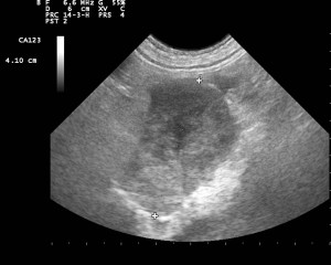
Fig.1 Mid-abdomen. An oval hypoechoic heterogeneous structure is seen between calipers.
This is an intra-abdominal lymph node. It is enlarged, rounded with a heterogeneous texture and diffuse reduced echogenicity. These features are more commonly seen in neoplastic lymphoadenopathy.
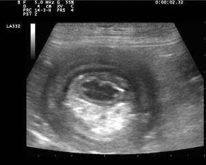
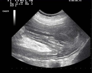
Fig. 2a & 2b above show the Mid-ventral abdomen. Sagittal and transverve view of the same area.
This is an intestinal intussuseption. Note the inner bowel loop (intussusceptum) showing a fairly normal wall; this is surrounded by hyperechoic mesenteric fat and by an outer intestinal loop (intussuscipiens) the wall of which is thickened and there is reduced layers demarcation.
At ultrasound examination the intestinal loop oral to the intussuception showed a thickened hypoechoic wall with loss of layers; liver and spleen were found to be enlarged and hyperechoic as well. Ultrasound guided fine needle aspiration biopsies were performed; samples from lymph nodes, liver and spleen were harvested.
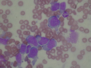
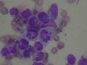
Figure 3a and 3b. show Cytological features of high-grade lymphoma.
Note that in figure 3b. there is two cells in mitoses.

