Focus on Orthopaedics: forelimb lameness in the young dog
Shoulder OCD
- Arthroscopy for diagnosis and treatment simultaneously in both shoulders
- Lameness resolved in >90% of dogs treated arthroscopically (Olivieri VCOT 2007)
Elbow Dysplasia: wide spectrum of disease
1. Medical coronoid disease is the most common
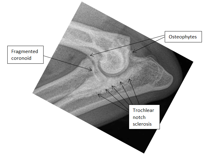
- Elbow fluoroscopic kinematics at Leipzig Vet School in August 2013 show that dysplastic dogs have substantial rotatory instability between the humerus and radio-ulnar joint cup. This abnormal motion is suggested as the cause of mechanical overload of the medial coronoid
- Wide spectrum of disease from fragmented medial coronoid to entire medial compartment syndrome (this is when there is complete cartilage loss of the medial humeral condyle and medial coronoid)
2. Osteochondrosis disecan of the medial humeral condyle
3. Un-united anconeal process
What can be done?
Early diagnosis is key (5-6 months onwards)
- Orthopaedic examination
- Radiographs x 3 views – flexed lateral, neutral medio-lateral, cranio-caual
Anthroscopy for diagnosis and treatment simultaneiously
- Minimally invasive
- Can go home the same day
- Bilateral examination
- “Shelbow” elbows and shoulders can be arthroscoped under the same anaesthetic if required

CT Scan
Further surgical treatment options include biceps ulnar release (BURP), ulnar osteotomies including bi-oblique dynamic proximal ulnar osteotomy, proximal abducting ulnar osteotomy (PAUL), resurfacing the humeral condyle (CUE) and elbow replacement or arthrodesis.
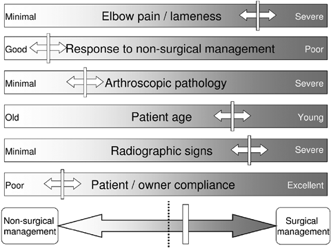
Management advice should always be given including exercise protocols, hydrotherapy, physiotherapy, diet and medical treatments including anti-inflammatories/analgesia

Don’t forget the differentials
- Panosteitis
- Metaphyseal osteopathy
- Juvenile septic arthropathy
- Incomplete ossification of the humeral condyle
- Trauma

