CNS disease in rabbits: E. cuniculi & the inner ear
Neurological disease in rabbits is common. Whilst undertaking a detailed neurological examination is difficult in rabbits, proprioceptive deficits, paresis and paralysis are readily detected. Indicators of spinal disease can include evidence of reduced (or lack of) grooming of the coat and perineum, perineal soiling and dermatitis. Diagnostic imaging and appropriate clinical pathology investigations should be carried out.
Grubber (2009) reports that 91% of CNS cases in rabbits were caused by E. cuniculi, the only real differential diagnoses being vestibular or inner ear disease – which may be differentiated by radiographic imaging of the bulla.
Even with the use of paired serology and IgG, together with IgM tests, the picture is confusing. Some 30% of rabbits have sub-clinical inner ear disease, which can cause similar concurrent signs.
| Sign | Inner ear | Brain stem (e.g. E. Cuniculi) |
|---|---|---|
| Head tilt | Present | Present |
| Circling | Present | Present |
| Falling, rolling | Present | Present |
| Nystagmus | Usually spontaneous and does not vary with head position | Usually not spontaneous - found with changes in head position |
| Direction of nystagmus | Usually horizontal, but can be rotatory | Usually positional, can be vertical or rarely, rotatory |
| Conscious proprioception | Normal | Delayed or absent |
| Horner's syndrome | May be present | Absent |
| Gait changes | Mild to severe ataxia | Ataxia and weakness |
| Postural reactions (hopping, hemi-walking, etc) | Normal if examined slowly | Weak or absent |
Rabbits with inner ear disease may present with lethargy, inappetence, pain, pruritis, swelling at the base of the ear, head tilt and facial paralysis; but most commonly these cases are sub-clinical.
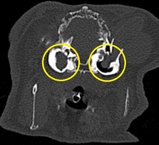
The aetiology is ascending Pasturella spp. infection via the eustachian tube rather than infection via the outer and middle ear. Until now bulla radiography has commonly been used diagnostically; however, we now know that its sensitivity as a diagnostic test is only 56%. The diagnostic test of choice is now CT scanning.

In relation to therapy of inner ear disease in rabbits, a surgical approach is mandatory. Many affected cases suffer bilateral disease.
Many anatomical and surgical texts are inaccurate in describing rabbits as having a vertical and horizontal ear canal similar to cats and dogs, as only a vertical canal is present check these guys out.
The vertical canal leads into the bony acoustic meatus, which then leads via the very narrow acoustic duct into the thick boned bulla. The bulla is 5mm deep, 7.5mm high and 11mm long. The lateral part of the ventral wall must be removed and the centre cleaned out.
The lining of the bulla and all its contents must be removed (and this should be sent for culture and sensitivity). Strict attention should be paid to decontamination, followed by surgical closure or marsupialisation and 6 weeks appropriate antibiosis.

